Preparation for prostatectomy
Patients in the pre-, peri- and post-operative period are under the care of an interdisciplinary team consisting of a surgeon an anaesthesiologist, a psychooncologist, physiotherapist and dietician as well as other specialty doctors depending on the overall health status. The necessary preparations include assessment of full blood count and blood smear and of kidney function as well as cross-matching.
Particular attention is given assessment made by anaesthesiologists and other specialist of patients with respiratory and respiratory diseases as well as those suffering from congestive heart failure and valvular insufficiency due to the increased risk of complications in their cases.
The anaesthesiologist starts perioperational antithrombotic therapy. Depending on the patient’s overall condition, they may also be assisted by other specialist in order to put in place the optimal pharmacological procedure.
The psychooncologist supports patients with cancers as well as their loved ones during the whole journey through the disease. They may also assist in minimising the adverse mental effects that the patient and his family experience in connection with the disease as well as in building a system of support to the patient, both internal (sense of worth, optimism, creativity and sense of humour) and external (family, significant others, friends and fellow workers). They also provides instruction on how to transform the attitude and thoughts that have a weakening effect on the patient and on reducing techniques, such as visualisations, relaxation or breathing exercises.
The physiotherapist works with the therapy and functional assessment of the pelvic floor and the overall condition of the patient. Physiotherapy used before and after the surgical intervention enables appropriate preparation for the procedure and maintaining its effects. Rehabilitated patients have a faster recovery rate with regard to the sexual function and urinary continence. Appropriate physiotherapy on the day of the procedure prevents complications in the form of pneumonia and venous thrombosis.
Of great importance for the recovery after cancerous diseases or extensive surgical interventions is correctly balanced nutrition. Therefore, the therapeutic team includes a dietician.
Procedure of removal of an ovary
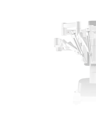
The procedure using the robot is performed under general anaesthesia. After administration of anaesthesia the patient is positioned on the operating table so as to avoid any injury during the procedure. During the procedure, the patient is in the so-called Trendelenburg position, i.e. with the feet elevated above the head. Then catheterisation of the patient is done. The catheter may remain in the bladder for the duration of the procedure or longer. Additionally, it may be necessary to use an intrauterine manipulator in order to obtain a better view of the pouch of Douglas.
At the beginning of each procedure, the so-called pneumoperitoneum is produced by filling the abdominal cavity with carbon dioxide in such a way as to obtain a convenient space in which to manipulate the instruments by elevating the abdominal walls from inside and pushing aside the intestines. Then the surgeon makes a few small abdominal incisions in order to introduce the optical system of the robot into the abdomen, which enables viewing the abdominal organs, as well as small-sided surgical instruments. On the screen in the console the surgeon gets a very high-definition colour image in considerable magnification. By means of some micro-tools the operator removes the ovary in its entirety. The removed lesions are placed in a special bag (so-called ‘endobag’) introduced into the abdomen and taken outside through one of the previously made incisions. Additionally, the surgeon performs a biopsy to take small samples of the tissue for further examination. After the procedure has been completed, the tools are removed from the abdomen and the carbon dioxide is released. The wounds are dressed using cosmetic sutures. During the procedure the abdominal cavity must be flushed from time to time in order to produce a good image of the operative field.
After the procedure, a drain may be inserted into the abdomen. After approx. 6 hours after the patient is already verticalised. Several hours after the surgery the patient may drink liquids and after the return of intestinal motility she can start eating easily digestible foods. The patient is discharged after about 2-3 days following the operation. After the surgery patients can return to their daily routine after about 5 days and to full activity after about two weeks.
The robotic method makes it possible to perform procedures without the necessity of opening the abdominal cavity, reduces postoperative pain ailments, promotes faster recovery, causes fewer postoperative wound infections and guarantees a better cosmetic effect.
Benefits for patients
The robotic system is a better tool, as compared with the classic or laparoscopic access, for resecting and removing lymph nodes while protecting the autonomic nerves during oncological procedures. Robot-assisted surgery enables surgical oncologists to perform minimally invasive, precise and complete procedures with good oncological clearance.
In comparison with procedures performed using the classical or partly laparoscopic methods, the use of a robot enables:
- collection of at least several tens of percent more lymph nodes
- smaller number of complications
- minimally invasive hysterectomy for large-sized uteri
- minimally invasive access in obese patients
- reduction of postoperative pain
- reduced blood loss
- better cosmetic effect
- shorter stay at hospital
- faster recovery
- possibility of operating on patients with a negative family history
- better cosmetic effect compared with the classic techniques

 +48 785 054 460


